
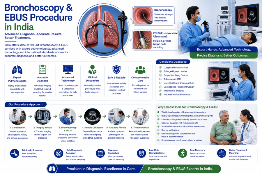
Bronchoscopy generally takes 20 to 40 minutes, while EBUS-TBNA usually takes 30 to 90 minutes, depending on the complexity of the procedure.
Most procedures are performed as day-care procedures, and patients are discharged on the same day. Selected patients may require 1 day of observation.
The procedure is usually performed under local anesthesia with intravenous sedation or short general anesthesia, depending on the patient's condition and the complexity of the procedure.
1,500 to 4,000 USD*
Bronchoscopy and Endobronchial Ultrasound (EBUS) are advanced minimally invasive procedures used to diagnose and, in selected cases, treat diseases affecting the lungs, airways, and surrounding lymph nodes. A bronchoscope is a thin, flexible tube equipped with a high-definition camera that is passed through the nose or mouth into the airways, allowing specialists to examine the respiratory tract, obtain tissue samples, remove foreign bodies, or treat airway blockages. EBUS combines bronchoscopy with real-time ultrasound imaging, enabling precise biopsy of lymph nodes and lung masses without the need for open surgery.
India has become one of the world's leading destinations for advanced bronchoscopy and EBUS procedures. Patients from Africa, the Middle East, and many other countries choose India because of its internationally accredited hospitals, experienced interventional pulmonologists, state-of-the-art bronchoscopy suites, advanced diagnostic technology, and affordable treatment costs compared to many developed countries.
Recent advancements in bronchoscopy have significantly improved diagnostic accuracy and patient safety. Leading hospitals in India now offer Endobronchial Ultrasound (EBUS-TBNA), Electromagnetic Navigation Bronchoscopy (ENB), Robotic-Assisted Bronchoscopy (selected centers), Cryobiopsy, Radial EBUS, Autofluorescence Bronchoscopy, Narrow Band Imaging (NBI), Therapeutic Bronchoscopy, Bronchial Stenting, Laser Airway Therapy, Argon Plasma Coagulation (APC), and AI-assisted Bronchoscopic Imaging. These technologies enable early diagnosis of lung cancer, tuberculosis, interstitial lung disease (ILD), infections, and mediastinal lymph node disorders while reducing the need for invasive surgical biopsies.
Whether you require evaluation for lung cancer, persistent cough, tuberculosis, enlarged lymph nodes, unexplained lung lesions, or airway obstruction, India offers world-class bronchoscopy and EBUS services with international standards of quality, safety, and personalized patient care.
Patients may require Bronchoscopy or EBUS if they experience:
Early evaluation helps identify serious lung diseases before they progress.
Bronchoscopy or EBUS may be recommended for patients with:
Before the procedure, patients undergo a complete respiratory assessment.
Pre-procedure investigations may include:
Patients are usually advised to fast for 6 to 8 hours before the procedure.
International patients can submit their medical reports online to receive an expert opinion before travelling to India.
Bronchoscopy and EBUS are performed by experienced interventional pulmonologists using advanced endoscopic equipment.
A thin flexible bronchoscope is gently inserted through the nose or mouth to examine the airways, trachea, and bronchi.
Real-time ultrasound attached to the bronchoscope allows visualization of lymph nodes and structures surrounding the airways.
A fine needle is passed through the airway wall under ultrasound guidance to obtain tissue samples from enlarged lymph nodes or lung masses for laboratory analysis.
Small tissue samples are collected from abnormal areas to diagnose lung cancer, infections, inflammatory diseases, or interstitial lung disorders.
A specialized cryoprobe freezes and removes larger lung tissue samples, improving the diagnosis of Interstitial Lung Disease (ILD).
Bronchoscopy can also be used to:
The most commonly performed procedure for diagnosing and treating airway diseases.
Used to evaluate lymph nodes and stage lung cancer without open surgery.
Provides detailed imaging of small peripheral lung lesions.
Advanced navigation technology helps access small lung nodules that are difficult to reach with conventional bronchoscopy.
Available at selected centers for highly accurate biopsy of small peripheral lung nodules.
Most patients recover quickly after bronchoscopy or EBUS.
Post-procedure care includes:
Most patients return home on the same day and resume normal daily activities within 24 hours. Mild sore throat or cough usually resolves within one or two days.
The biopsy results obtained during bronchoscopy or EBUS help specialists develop a personalized treatment plan for conditions such as lung cancer, tuberculosis, pulmonary fibrosis, sarcoidosis, and other respiratory diseases. Early diagnosis enables timely treatment and improves long-term outcomes.
Patients may require Bronchoscopy or EBUS if they experience:
Early evaluation helps identify serious lung diseases before they progress.
Bronchoscopy or EBUS may be recommended for patients with:
Before the procedure, patients undergo a complete respiratory assessment.
Pre-procedure investigations may include:
Patients are usually advised to fast for 6 to 8 hours before the procedure.
International patients can submit their medical reports online to receive an expert opinion before travelling to India.
Bronchoscopy and EBUS are performed by experienced interventional pulmonologists using advanced endoscopic equipment.
A thin flexible bronchoscope is gently inserted through the nose or mouth to examine the airways, trachea, and bronchi.
Real-time ultrasound attached to the bronchoscope allows visualization of lymph nodes and structures surrounding the airways.
A fine needle is passed through the airway wall under ultrasound guidance to obtain tissue samples from enlarged lymph nodes or lung masses for laboratory analysis.
Small tissue samples are collected from abnormal areas to diagnose lung cancer, infections, inflammatory diseases, or interstitial lung disorders.
A specialized cryoprobe freezes and removes larger lung tissue samples, improving the diagnosis of Interstitial Lung Disease (ILD).
Bronchoscopy can also be used to:
The most commonly performed procedure for diagnosing and treating airway diseases.
Used to evaluate lymph nodes and stage lung cancer without open surgery.
Provides detailed imaging of small peripheral lung lesions.
Advanced navigation technology helps access small lung nodules that are difficult to reach with conventional bronchoscopy.
Available at selected centers for highly accurate biopsy of small peripheral lung nodules.
Most patients recover quickly after bronchoscopy or EBUS.
Post-procedure care includes:
Most patients return home on the same day and resume normal daily activities within 24 hours. Mild sore throat or cough usually resolves within one or two days.
The biopsy results obtained during bronchoscopy or EBUS help specialists develop a personalized treatment plan for conditions such as lung cancer, tuberculosis, pulmonary fibrosis, sarcoidosis, and other respiratory diseases. Early diagnosis enables timely treatment and improves long-term outcomes.
Yes. Bronchoscopy and EBUS procedures in India are significantly more affordable than in many developed countries while maintaining international standards of respiratory care.
Bronchoscopy allows direct visualization of the airways, while EBUS combines bronchoscopy with ultrasound imaging to safely biopsy lymph nodes and lung masses located outside the airways.
Recent advancements include Robotic Bronchoscopy, EBUS-TBNA, Electromagnetic Navigation Bronchoscopy (ENB), Cryobiopsy, Radial EBUS, Narrow Band Imaging (NBI), AI-assisted imaging, and advanced therapeutic bronchoscopy.
No. The procedure is usually performed under local anesthesia with sedation or short general anesthesia, and most patients experience minimal discomfort.
Yes. International patients receive comprehensive assistance including online consultations, medical visas, accommodation, airport transfers, interpreter services, and follow-up care.
Most pathology reports are available within 3 to 7 working days, although specialized molecular or microbiological tests may require additional time.
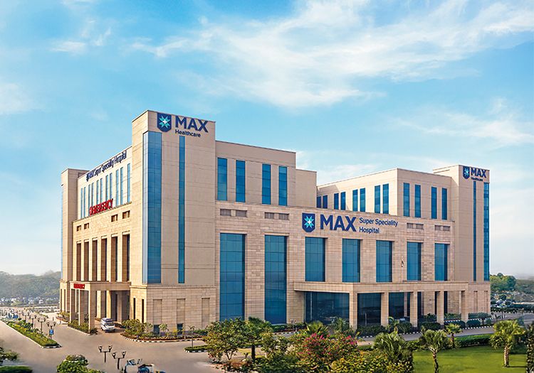
Jaypee Hospital Road, Goberdhanpur, Sector 128, Noida, Uttar Pradesh – 201304, India
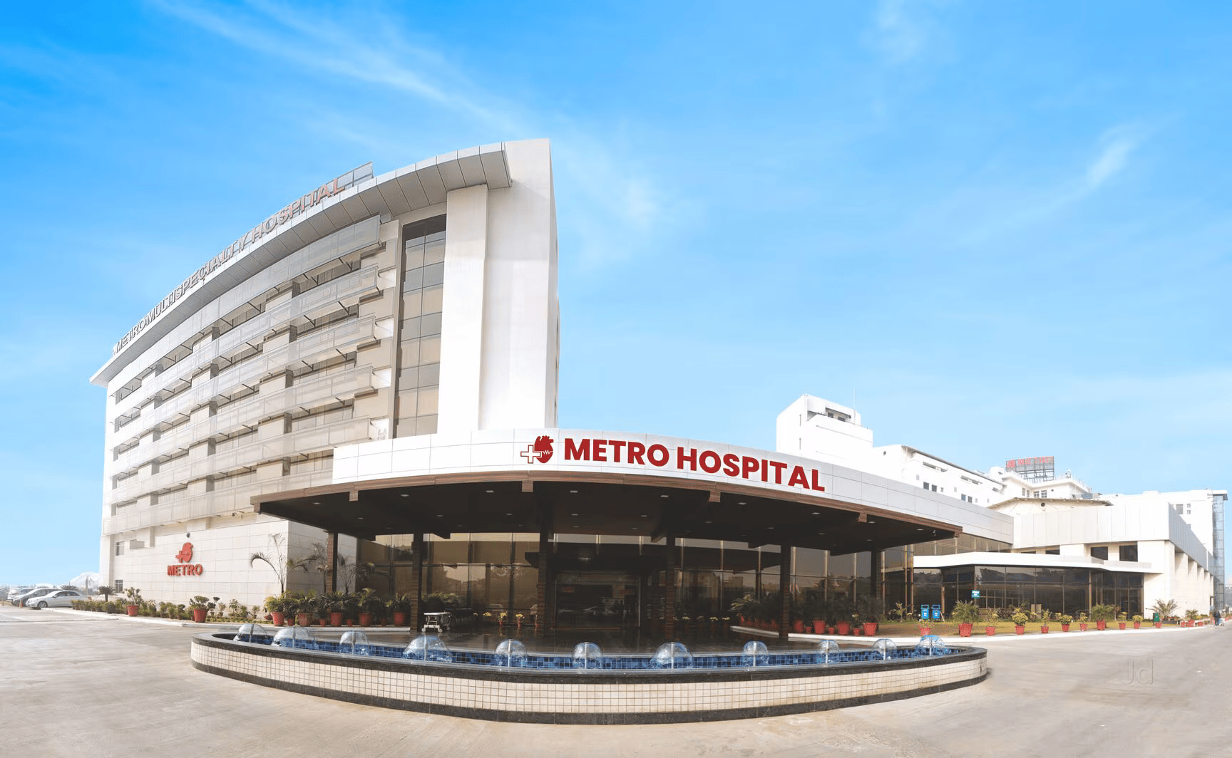
Sector 16A, Faridabad, Haryana – 121002, India
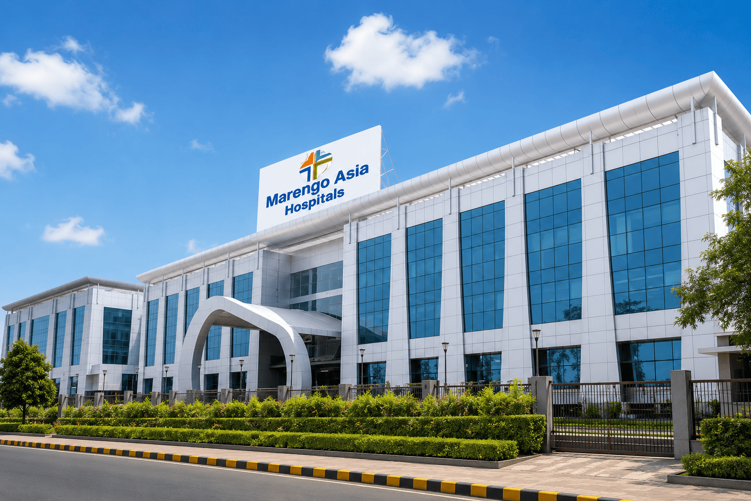
Plot No. 1, HUDA Staff Colony, Sector 16, Faridabad, Haryana – 121002, India
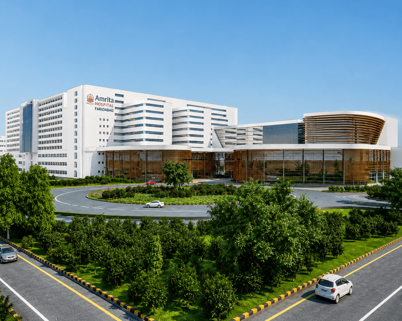
Mata Amritanandamayi Marg, Sector 88, Faridabad, Haryana – 121014, India
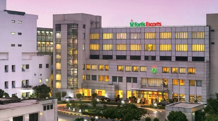
Okhla Road, Opposite Sukhdev Vihar Metro Station, New Delhi – 110025, India
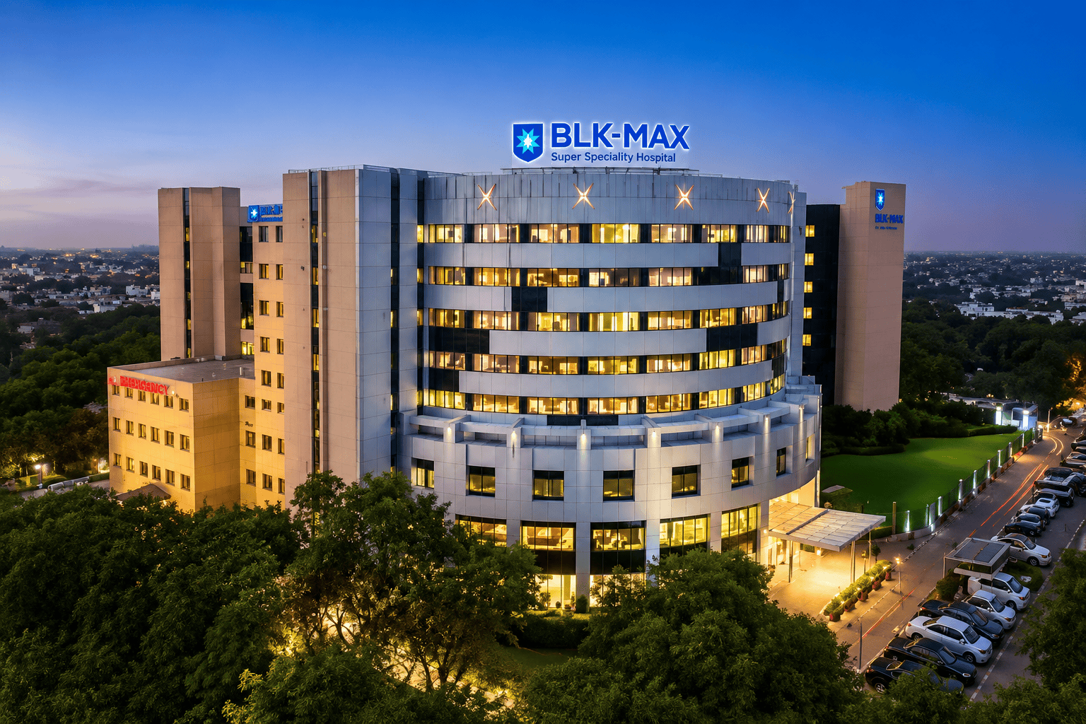
Pusa Road, Rajendra Place, New Delhi – 110005, India
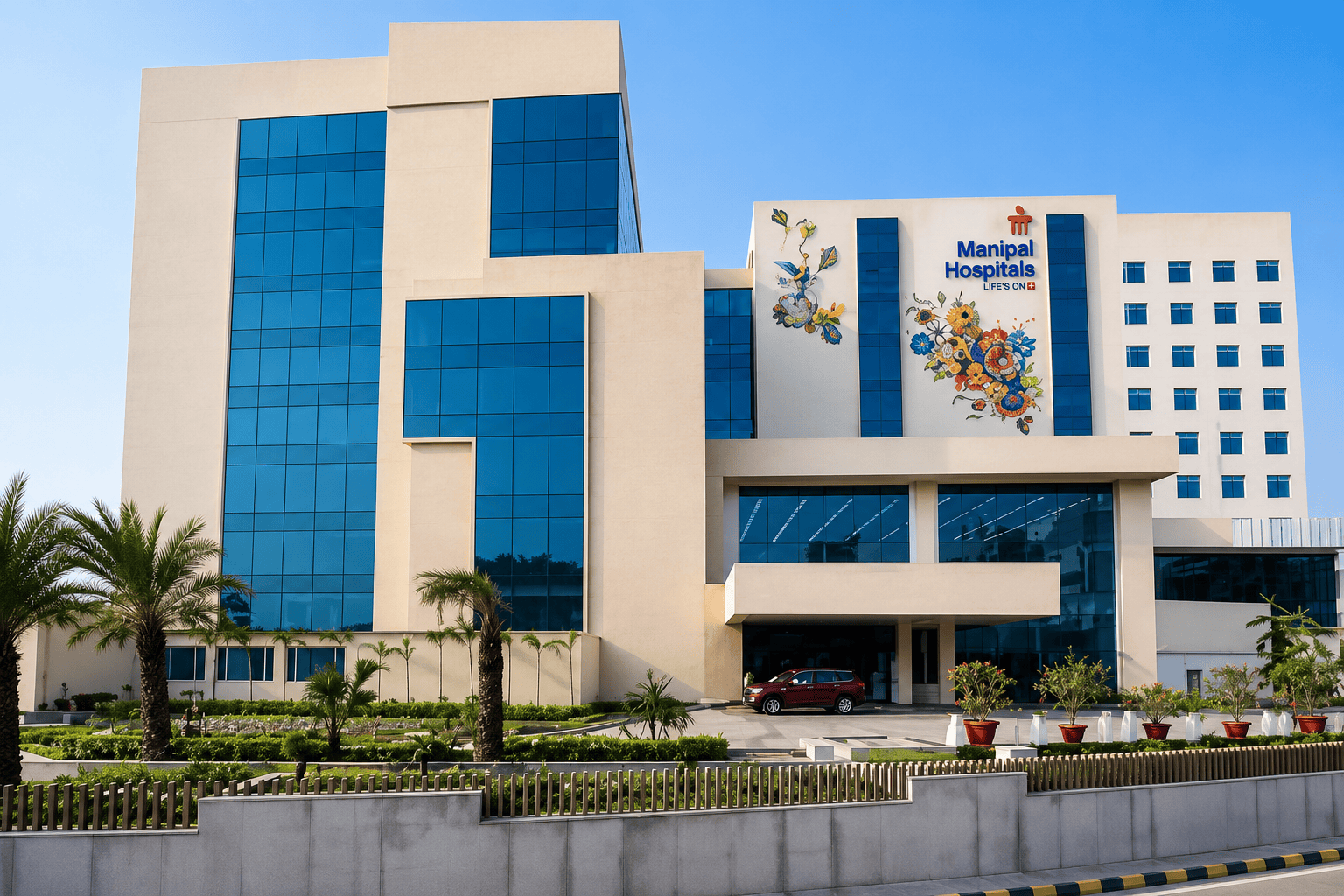
Palam Vihar, Sector 6, Dwarka, New Delhi – 110075, India
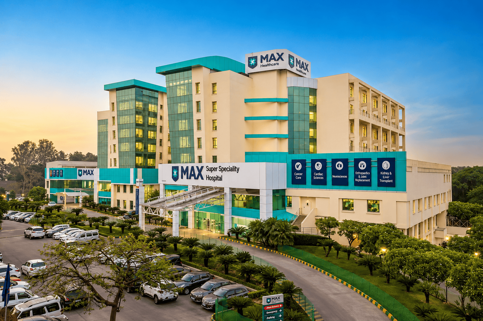
1 & 2, Press Enclave Road, Saket Institutional Area, Saket, New Delhi – 110017, India
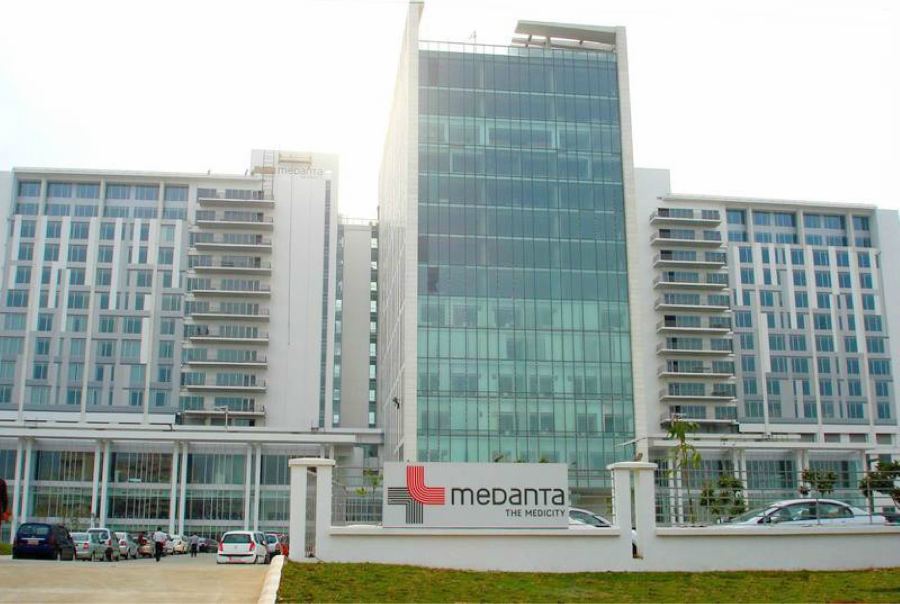
CH Baktawar Singh Road, Sector 38, Gurugram, Haryana – 122001, India
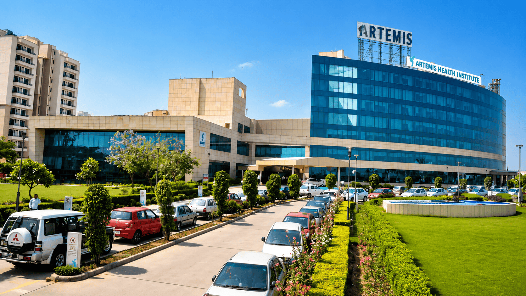
Sector 51, Gurugram, Haryana – 122018, India
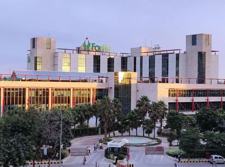
Sector - 44, Gurugram, Haryana – 122002, India
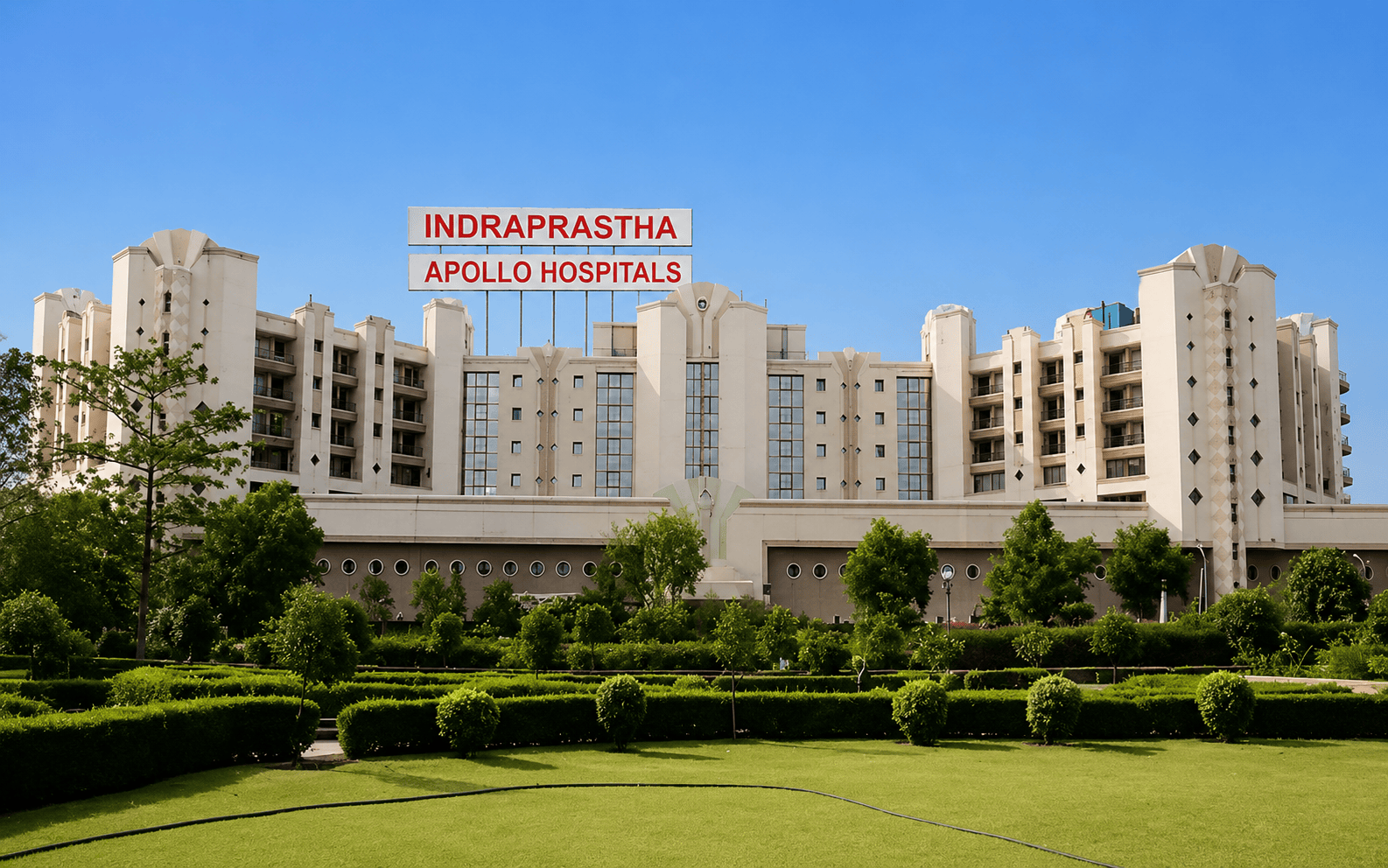
Sarita Vihar, Delhi-Mathura Road, New Delhi – 110076, India